Home » Without Label » Long Bone Diagram Labled / Label Skeletal Long Bone Diagram Quizlet : The diaphysis is the tubular shaft that runs between the proximal and distal ends of the bone.
Long Bone Diagram Labled / Label Skeletal Long Bone Diagram Quizlet : The diaphysis is the tubular shaft that runs between the proximal and distal ends of the bone.
Long Bone Diagram Labled / Label Skeletal Long Bone Diagram Quizlet : The diaphysis is the tubular shaft that runs between the proximal and distal ends of the bone.. Altogether, the skeleton makes up about 20 percent of a person's body weight. You should make a label that represents your brand and creativity, at the same time you shouldn't forget. In the diagram above, you can see all of the bones of the foot clearly labeled. Parts of long bone (applies to other bones too). The blood vessels inside a bone.
A labeled diagram of a long bone. This is an online quiz called label a long bone. The ends of long bones are called. They are one of five types of bones: Designed to fit the bone or bones it attaches to.
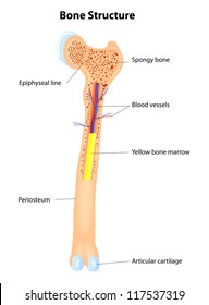
Skeletal System 1 The Anatomy And Physiology Of Bones Nursing Times from cdn.ps.emap.com Long bones are the most common bones found in the human body. The shiny, articulating cartilage on the ends of a bone. Label number 1 in the diagram indicates which part of the bone? Bone · august 7, 2016. Start studying long bone labeled. A = epiphysis b = diaphysis c = articular cartilage d = periosteum f = compact bone g = medullary cavity (yellow marrow) h = endosteum j = epiphyseal line (growth plate) coloring worksheet for this image. A typical long bone showing gross anatomical features. The tough membrane covering the shaft of the bone.
They are composed mostly of compact bone, and are roughly cylindrical in shape with enlarged ends filled with spongy bone.
Next to the tibia is the fibula, the thinner, weaker bone of the lower leg. You should make a label that represents your brand and creativity, at the same time you shouldn't forget. The long bones are those that are longer than they are wide. The blood vessels inside a bone. Human skeleton with each bone name 12 photos of the human skeleton with each bone name human skeleton with bone names, bone, human skeleton with bone names. Bone · august 7, 2016. Start studying long bone labeled. Anatomy of a long bone. Labeled diagram of an osteon. What do we mean by an 'articulation'? The membrane lining the bone cavity. The outside of the flat bone consists of a layer of connective tissue called the periosteum. A = epiphysis b = diaphysis c = articular cartilage d = periosteum f = compact bone g = medullary cavity (yellow marrow) h = endosteum j = epiphyseal line (growth plate) coloring worksheet for this image.
It can be found under the periosteum and in the diaphyses of long bones, where it provides support and protection. There is a printable worksheet available for download here so you can take the quiz with pen and paper. Red bone marrow fills the spaces between the spongy bone in some long bones. The tough membrane covering the shaft of the bone. Bone · august 7, 2016.
Long Bone Anatomy Images Stock Photos Vectors Shutterstock from image.shutterstock.com As part of your level 2 anatomy and physiology exam, you need to be aware of the structure of a long bone and know. Diagram of the femur (thigh bone) on the right, notice Compact bone is the denser, stronger of the two types of bone tissue ( (figure) ). A labeled diagram of a long bone. It can be found under the periosteum and in the diaphyses of long bones, where it provides support and protection. A long bone has two parts: In the diagram above, you can see all of the bones of the foot clearly labeled. Compact bone is the denser, stronger of the two types of bone tissue ( link ).
An easy and convenient way to make label is to generate some ideas first.
Labeling portions of a long bone. It is also known as the calf bone, as it. Bone · august 7, 2016. It contains few spaces and provides protection and. In the diagram above, you can see all of the bones of the foot clearly labeled. Anatomy of a long bone. Each epiphysis is shaped differently; The ends of a long bone contain spongy bone and an epiphyseal line. Next to the tibia is the fibula, the thinner, weaker bone of the lower leg. A long bone has a shaft and 2 ends. You should make a label that represents your brand and creativity, at the same time you shouldn't forget. The structure of a long bone: A long bone has two parts:
As part of your level 2 anatomy and physiology exam, you need to be aware of the structure of a long bone and know. Altogether, the skeleton makes up about 20 percent of a person's body weight. The shiny, articulating cartilage on the ends of a bone. Bones are classified by their shape—as long, short, flat, and irregular. This is an online quiz called label the long bone there is a printable worksheet available for download here so you can take the quiz with pen and paper.
Femur Bone Anatomy Labeled Diagram Quiz Color Coded Parts Skeletal System Lower Extremity Ezmed from images.squarespace-cdn.com The ends of a long bone contain spongy bone and an epiphyseal line. Damaged joint and healthy joint. Diagram of the femur (thigh bone) on the right, notice This is an online quiz called label the long bone there is a printable worksheet available for download here so you can take the quiz with pen and paper. What do we mean by an 'articulation'? Long bones are those that are longer than they are wide. The membrane lining the bone cavity. A long bone has two parts:
There is a printable worksheet available for download here so you can take the quiz with pen and paper.
The diaphysis and the epiphysis. Start studying long bone labeled. Long, short, flat, irregular and sesamoid.long bones, especially the femur and tibia, are subjected to most of the load during daily activities and they are crucial for skeletal mobility.they grow primarily by elongation of the diaphysis, with an epiphysis at each end of the growing bone. A long bone is a. Parts of long bone (applies to other bones too). Human skeleton with each bone name. There is a printable worksheet available for download here so you can take the quiz with pen and paper. In these labeled examples, a human femur is represented without identifying many of the unique characteristics that help differentiate the femur bone from other bones in the human body. Used figure 6.2 in book. They are one of five types of bones: The long bones are those that are longer than they are wide. A = epiphysis b = diaphysis c = articular cartilage d = periosteum f = compact bone g = medullary cavity (yellow marrow) h = endosteum j = epiphyseal line (growth plate) coloring worksheet for this image. Next to the tibia is the fibula, the thinner, weaker bone of the lower leg.